Back Of Skull Anatomy : Skull, posterior view with labels - Axial Skeleton Visual ... / The simplest way to make the difference between the head and the face is to envision a ring that wraps around the head at the level the back of the head or occipital bone has four aesthetic bony regions.
Back Of Skull Anatomy : Skull, posterior view with labels - Axial Skeleton Visual ... / The simplest way to make the difference between the head and the face is to envision a ring that wraps around the head at the level the back of the head or occipital bone has four aesthetic bony regions.. Their number and location vary. Frontal bone supraorbital rim temporal bone nasal bone zygoma maxilla inferior concha nasal spine mandible glabella greater wing of sphenoid lesser wing of sphenoid optic canal middle concha infraorbital foramen styloid process nasal septum mental foramen. Upon reaching maturity, our skull bones fuse to produce a rigid protective shell for the soft nervous. In order to be light, the skull is made up by flat and irregular bones, and has hollow spaces called the sinuses. The skull bones can be classified into two groups:
The greater portion of the anterior floor is convex and the most important anatomic structures below the anterior cranial fossa are the orbits and the paranasal sinuses. The cranium (skull) is the skeletal structure of the head that supports the face and protects the brain. This anatomic region is complex and poses surgical challenges for otolaryngologists and neurosurgeons alike. The skull bones can be classified into two groups: « back show on map ».
131 best MY skull anatomy images on Pinterest | Bones ... from i.pinimg.com The skull performs vital functions. The cranial vault denotes the top, sides, front, and back of the cranium. Learn more about the anatomy and function of the skull in humans and other vertebrates. Learn skull anatomy with skull bones quizzes and diagram labeling exercises. Learn about skull base anatomy with free interactive flashcards. « back show on map ». Human skull from the front. The frontal, parietal, temporal and occipital bones are joined at the cranial sutures.
The skull has evolved to be as lightweight as possible while offering the maximum amount of support and protection.
Protects the skull while permitting growth. Upon reaching maturity, our skull bones fuse to produce a rigid protective shell for the soft nervous. Foramina inside the body of humans and other animals. Learn about skull base anatomy with free interactive flashcards. The skull has a single occipital condyle.7 the skull consists of five major bones: Learn skull anatomy with skull bones quizzes and diagram labeling exercises. Provides bony architecture to major neurovascular and lymphatic structures. « back show on map ». Human anatomy for muscle, reproductive, and skeleton. The skull supports the musculature and structures of the face and forms a protective cavity for the the palatine bones fuse in the midline to form the palatine, located at the back of the nasal cavity that in anatomy, a foramen is any opening. Looking at it from the inside it can be subdivided into. Inferior view of base of the skull. Norma basalis ( anterior part , middle part and posterior part ).
Skull anatomy | with labels. The sagittal suture is the line where the right and left parietal bone are in contact. Excluding ear ossicles, it is made of 22 bones. It offers protection to the brain, eye balls, inner ears, and nasal passages. Inferior view of base of the skull.
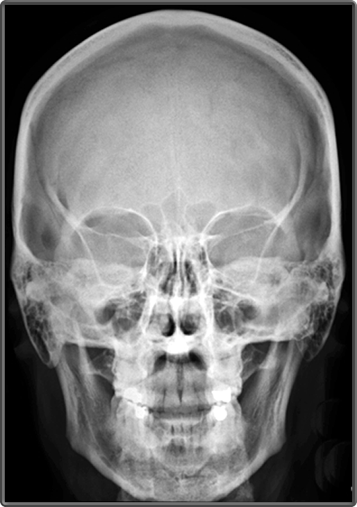
Clinical Anatomy | Radiology | Skull Sinuses from www.clinicalanatomy.ca A thorough description is beyond the. This anatomic region is complex and poses surgical challenges for otolaryngologists and neurosurgeons alike. The upper back is a complex area containing a number of muscles that perform various actions on the scapulae shoulder blades and humerus. The cranium (skull) is the skeletal structure of the head that supports the face and protects the brain. Anatomical structures of the skull include: Excluding ear ossicles, it is made of 22 bones. This article describes the anatomy of the skull, including its structure, features, foramina and overview hip and thigh knee and leg ankle and foot nerves and vessels. Protects the skull while permitting growth.
Skull bones aren't fused together at birth.
Foramina inside the body of humans and other animals. Human anatomy for muscle, reproductive, and skeleton. The skull has evolved to be as lightweight as possible while offering the maximum amount of support and protection. The cranium (skull) is the skeletal structure of the head that supports the face and protects the brain. A thorough description is beyond the. It is comprised of many bones, formed by intramembranous ossification, which are joined together by sutures (fibrous joints). Bone of back of skull. Learn more about the anatomy and function of the skull in humans and other vertebrates. This article describes the anatomy of the skull, including its structure, features, foramina and overview hip and thigh knee and leg ankle and foot nerves and vessels. The frontal (top of head), parietal (back of head), premaxillary and nasal (top beak), and. Skull anatomy | with labels. The skull base is the inferior portion of the neurocranium. « back show on map ».
So, the human skull consists of 23 bones. The skull bones can be classified into two groups: The sagittal suture is the line where the right and left parietal bone are in contact. It supports and protects the face and the brain. Upon reaching maturity, our skull bones fuse to produce a rigid protective shell for the soft nervous.
Human anatomy videos: Skull base part 1 - YouTube from i.ytimg.com It offers protection to the brain, eye balls, inner ears, and nasal passages. Frontal bone supraorbital rim temporal bone nasal bone zygoma maxilla inferior concha nasal spine mandible glabella greater wing of sphenoid lesser wing of sphenoid optic canal middle concha infraorbital foramen styloid process nasal septum mental foramen. Skull anatomy | with labels. During childhood development, the skull bones remain somewhat separated, allowing for growth of the brain and skull. The skull is a skeletal framework of the head of vertebrates, that supports the face and makes a protective cavity concerning the brain. So, the human skull consists of 23 bones. The bbc is not responsible for the content of external websites. In order to be light, the skull is made up by flat and irregular bones, and has hollow spaces called the sinuses.
The skull base is the inferior portion of the neurocranium.
Norma basalis ( anterior part , middle part and posterior part ). Related posts of bone of back of skull. The skull begins to form prior to week 12 of embryogenesis. It supports and protects the face and the brain. Learn about skull base anatomy with free interactive flashcards. The upper back is a complex area containing a number of muscles that perform various actions on the scapulae shoulder blades and humerus. Ct anatomy of skull, axial reconstruction, bone window. The frontal, parietal, temporal and occipital bones are joined at the cranial sutures. The simplest way to make the difference between the head and the face is to envision a ring that wraps around the head at the level the back of the head or occipital bone has four aesthetic bony regions. These joints fuse together in adulthood. Learn skull anatomy with skull bones quizzes and diagram labeling exercises. William is a final year medical student in australia who has taught anatomy to tertiary science and. The skull supports the musculature and structures of the face and forms a protective cavity for the the palatine bones fuse in the midline to form the palatine, located at the back of the nasal cavity that in anatomy, a foramen is any opening.


0 Komentar